Alberto Diaspro and Giuseppe Sito
https://www.giuseppesito.it/nuovo/wp-content/uploads/2025/03/surgeries-06-00021.pdf
Background: The attractiveness of the central area (the so-called mid-face area or middle third) has a strong impact on the observer, and the treatment of aging in this area is therefore considered a key component in facial rejuvenation. A standardized photographic and three-dimensional analysis was conducted in this observational study to determine the outcome of volumetric restoration procedures of the mid-face area with HA injection, providing an objective, repetitive, and reliable evaluation of this facial rejuvenation technique. Methods: In total, 47 patients were treated with two types of HAbased dermal fillers, and calibrated, stereoscopic images of the face were taken with volume reconstruction and analysis software performed before (t0), 45 days after HA implantation (t1), and at the check-up after the end of follow-up (t2). Results: In total, 39 out of 47 patients completed the study, which showed an overall volume restoration of 4.46 ± 1.34 mL at
45 days (t0–t1) after HA implantation, maintaining a value of 1.23 ± 0.68 mL at the end of the 318-day follow-up (t0–t2). Conclusions: The results of this study indicate that rejuvenation of the mid-facial region through volumetric restoration with an HA filler leads to an indirect volumetric effect that is clinically more significant than the actual injected volume and equally long-lasting.
Keywords: mid-face; hyaluronic acid; volumetric; rejuvenation
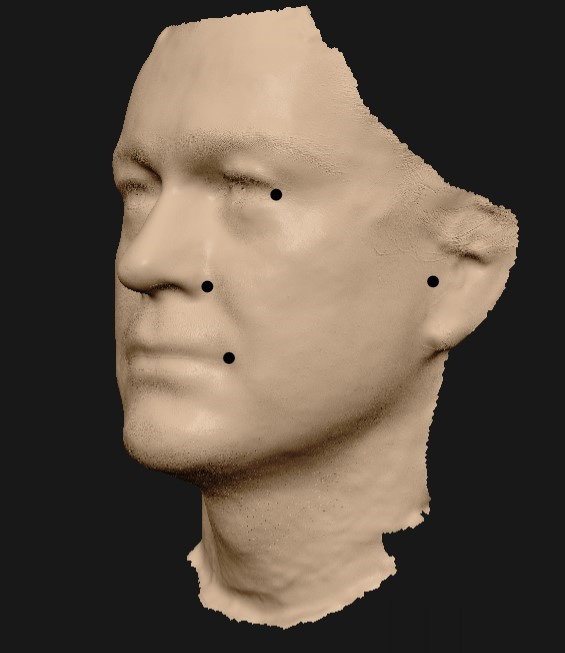
Figure 1. Reference points: pre-tragal (point Ar—Articularis), lateral canthus (point Ex—Exocanthon), alar cartilage (point Al—Alare), and oral commissure (point Ch—Cheilon).
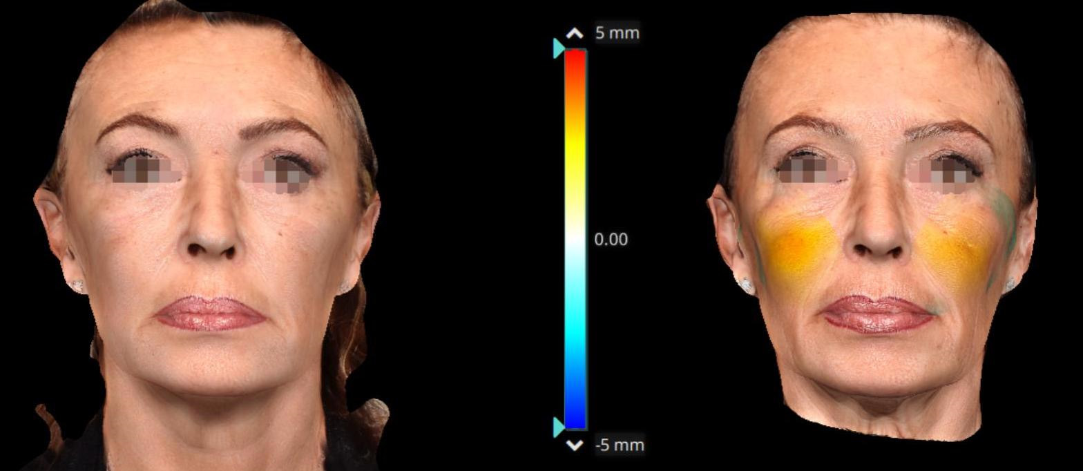
Figure 2. Frontal view with color scale, after and before injection therapy—patient 1.
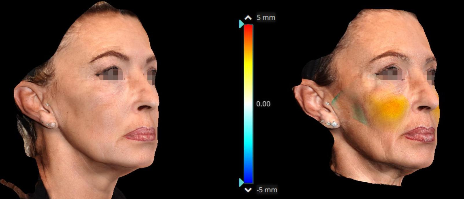
Figure 3. Oblique view with color scale, after and before injection therapy—patient 1.
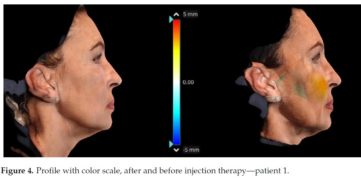
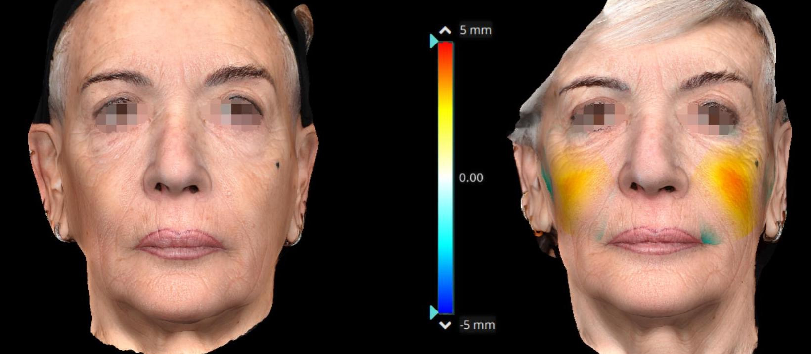
Figure 5. Frontal view with color scale, after and before injection therapy—patient 2.
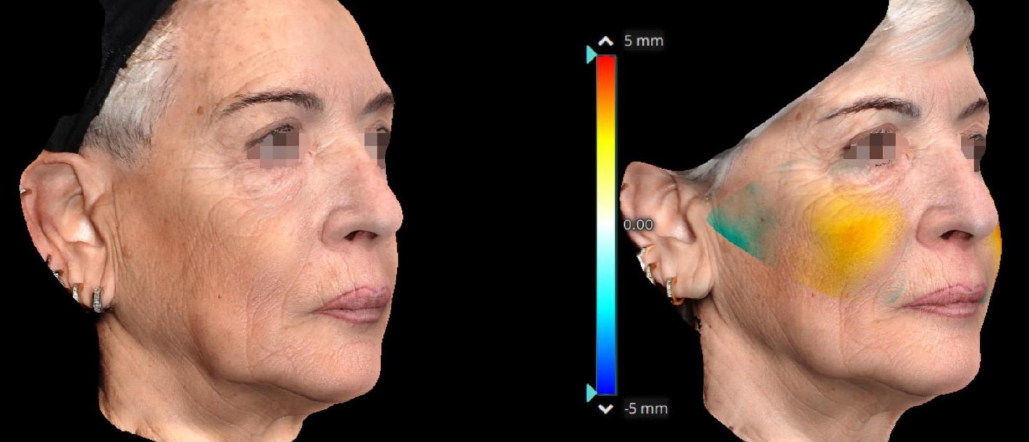
Figure 6. Oblique view with color scale, after and before injection therapy—patient 2.
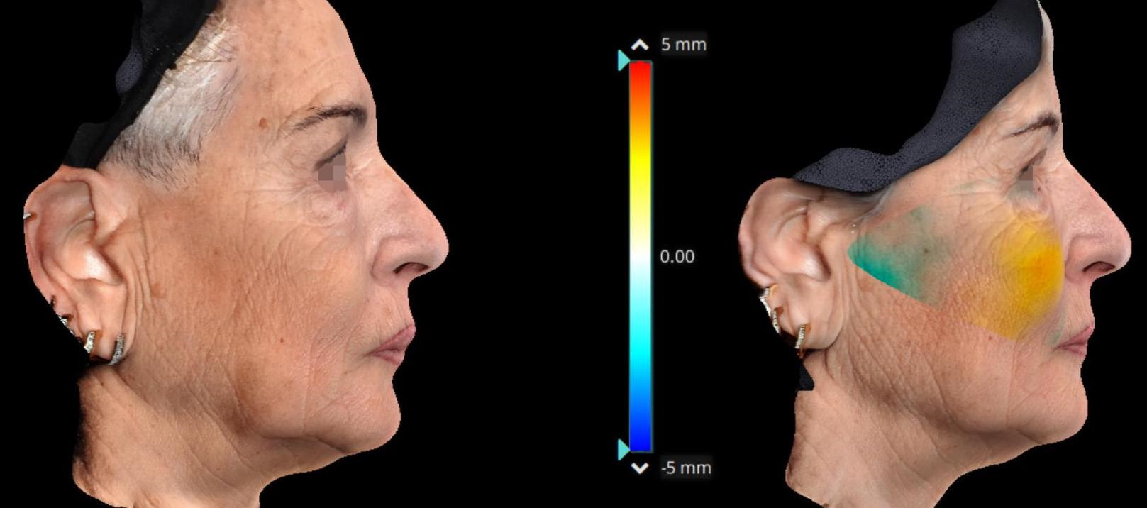
Figure 7. Profile with color scale, after and before injection therapy—patient 2.
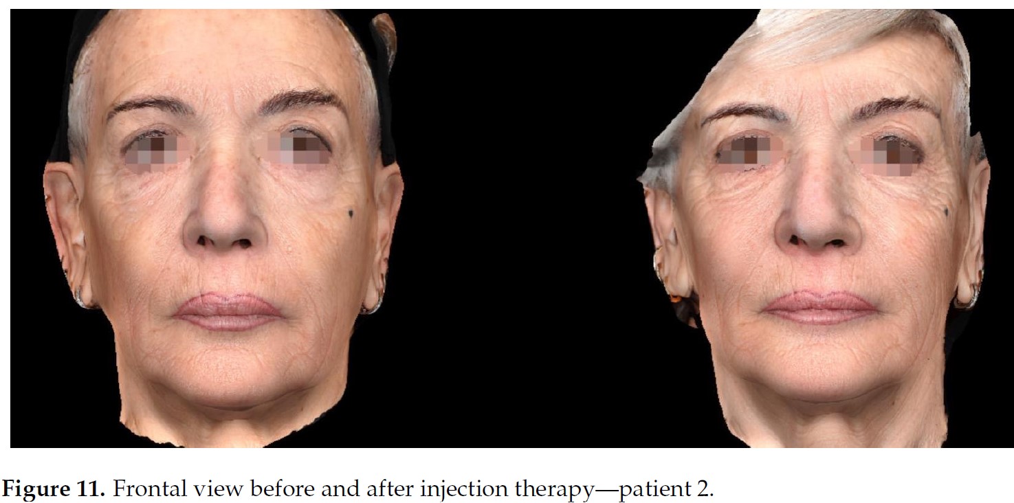
Figure 8. Frontal view before and after injection therapy—patient
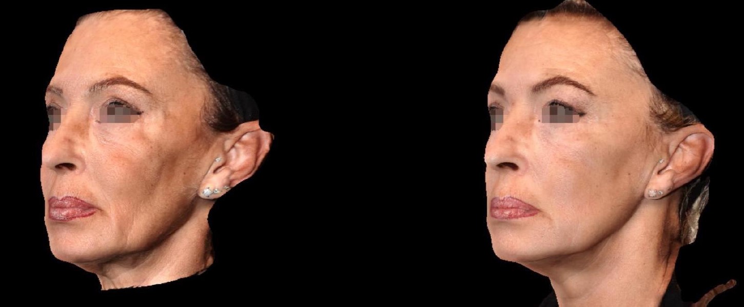
Figure 9. Oblique view before and after injection therapy—patient 1